


World War I
World War I had a huge impact on the world as we know it, including the field of medicine. From 1914 to 1918, normal life at that time was paused as soldiers fought for their lives to defend their country. The war led to mass death and destruction, and for the first time something else was a bigger killer than disease. However, in the same way that Ambroise Paré learned from his time working on the battlefield, the war brought with it complex injuries which forced people to consider new treatments and develop new ideas.
The Western Front
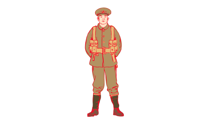
Until now, when a war broke out it would usually mean troops of soldiers would advance across large stretches of land to attack the enemy and force them to retreat or surrender. In World War I, this wasn’t the case. In Western Europe, there was very little movement and often periods of stalemate. Britain, France and Belgium fought Germany at the Western Front. When Germany began digging long ditches called trenches in France and Belgium, Britain and France had no choice but to build trenches opposite them. This created an area called “No Man’s Land” in between, which soldiers had to cross to attack the opposite trenches. This kind of warfare led to terrible injuries and huge numbers of deaths.
The Trenches
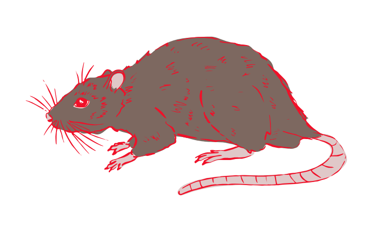
The conditions in the trenches were miserable and unhygienic, not to mention dangerous! When soldiers weren’t under enemy fire and fighting for their life, they’d be trying to avoid rats and sinking in the mud. To try and prevent soldiers’ feet sinking, duckboards were built using planks of wood. There was some shelter available in the dugouts, but otherwise the trenches were out in the open whatever the weather (including lots of rain!).
War Wounds and Diseases
Soldiers fighting during World War One were vulnerable to lots of awful injuries, as well as illness and disease.
Gunshot wounds
The range of weaponry used during the war came with a range of terrible injuries, some of them deadly. When shot, rifles and machine guns would pierce organs and break bones and led to thousands of soldiers needing amputations. If they didn’t die straight away from their injuries, soldiers were likely to bleed out and experience organ failure.
Shrapnel wounds
Shrapnel shells were projectiles filled with tiny metal balls that would explode, and hot fragments of the shell’s casing would hit soldiers, ripping through their skin or lodging inside their body. They were responsible for over 50% of casualties during World War I. Because of their deadly impact, especially if they hit a soldier in the face or head, metal helmets were given to soldiers to try and protect them better. You can see an example of shell fragments from Surgeon’s Hall Museum in our Source Gallery.
Gas
Poison gas was introduced to the war by Germany in 1915. Chlorine gas would strip away the lung lining and soldiers would drown from liquid in their lungs. Phosphene gas worked slowly over 48 hours, causing soldiers to vomit and spasm while their lungs drowned in yellow liquid. Mustard gas (although less deadly) caused burning and swelling of the skin and eyes, often leading to blindness. Fortunately, by 1916, gas masks prevented the majority of gas-related injuries and deaths. Unfortunately, however, those who were attacked by poison gas would take up spaces when soldiers with life-threatening injuries needed to be treated.
Shell shock
At the time World War I began, we didn’t understand mental health as clearly as we do today. The horrors of the battlefield led to many soldiers suffering from what we now call Post-Traumatic Stress Disorder (or PTSD). At the time, over 80,000 British soldiers on the Western Front suffered from this condition and it was known as “shell shock”.
Patients would experience a range of physical and mental symptoms, from headaches and paralysis to flashbacks and memory loss. Originally, soldiers presenting with these symptoms were labelled as cowards, and many were executed as a result. However, as more cases occurred, the British Army began responding. Most patients were provided with rest and food, and a medical officer would talk to them about their experiences to try and persuade them to return to their post. Once shell shock was accepted as a genuine condition, patients were sent home or to hospital. During this time, new talking treatments were developed by a psychologist called William Rivers and patients were encouraged to take part in activities to improve their mental health. These ideas lasted beyond the war and have since been used for treating a range of mental health conditions.
Infection
Because of the muddy conditions, most war wounds became infected. Even if a soldier only suffered a mild injury, all it would take is for dirt to enter a wound and it could become deadly. Gangrene was the deadliest of the infections at this time – it would turn wounds green and fill them with gas, which was as bad as it sounds!
Trench foot
Because the trenches were full of thick mud and water, soldiers were constantly at risk of trench foot. When trench foot set in, a soldier’s toes would become numb and swollen, and would even turn blue. This could then lead to gangrene, which usually spread around the body.
Frostbite
Frostbite was another risk for soldiers’ feet, and hands too. It would cut off the circulation, and meant that many soldiers had to have their fingers or toes amputated. As the name suggests, frostbite attacked when soldiers were out in extremely cold temperatures.
Trench fever
Lice were a real problem for soldiers on the Western Front. They lived in soldiers’ skin and clothes, and their bites would leave behind itchy and painful blisters. If these bite wounds became infected, a soldier might develop pyrexia, which was known as trench fever. Its symptoms only lasted a matter of days, but included a fever, headaches and painful joints. This meant soldiers with trench fever weren’t fit enough to serve. The army tried everything to get rid of the lice, from lice repellent to fumigating uniforms, but they thrived in the trenches throughout the war.
Did you know?
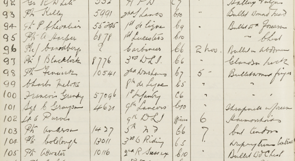
During World War I, some of Newcastle University’s buildings were used to form a hospital that treated wounded soldiers. Professor Frederick Pybus, who was a surgeon, carried out over 1,000 operations on soldiers who were brought here. We can see from his logbook that many of them were for gunshot and shrapnel wounds.
The Chain of Evacuation
For soldiers who had sustained serious injuries, it wasn’t possible for treatment to be given on the Front. This meant medical officers had to decide which evacuation route would be best for each case. The chain included:
- Stretcher bearers, who would take soldiers to safety on a stretcher. They often collected wounded soldiers at nighttime, and would do what they could to make sure the soldier was comfortable and carry out First Aid. They had access to basic supplies, like painkillers and bandages.
- Regimental Aid Posts (RAP), which is where stretcher bearers would often take soldiers. RAPs were set up right behind the front line, so weren’t the safest place to be. Here, medical officers would try to treat a soldier so they could return straight back to the front line. If this wasn’t possible, they’d be sent on to an Advanced Dressing Station.
- Advanced Dressing Stations (ADS), which were much further back from the front line. ADS had more protection than RAPs, and were often set up inside old, abandoned buildings or dugouts. Here, soldiers could receive further treatment at a basic level. If their injury needed more attention, they would be collected by an ambulance.
- Ambulances, which took soldiers from Advanced Dressing Stations to a Casualty Clearing Station (CCS). During the war, ambulances weren’t kitted out like they are today – it would be a bumpy ride from the ADS to the CCS!
- Casualty Clearing Stations (CCS), which were set up several miles away from the front line in sturdier spaces, like village halls and schools. At the CCS, a soldier would go through the triage process to determine what to do next. Those who could be saved would be treated immediately, but those who had suffered deadly injuries would be kept as comfortable as possible until they died. CCSs saw over 200 wounded soldiers a day, and usually didn’t have enough staff.
- Base hospitals, which could hold thousands of soldiers and would usually be based in an actual hospital. This was the last chance for a soldier to be treated before being sent home. Base Hospitals were set up by the coast and often near railway lines so soldiers could be brought there by the hospital trains. They had separate wards for different kinds of injuries.
The Royal Army Medical Corps and Queen Alexandra’s Imperial Military Nursing Service
From the late nineteenth century, any medical personnel who cared for soldiers’ health became a member of The Royal Army Medical Corp (RAMC). From doctors and dentists to ambulance drivers and opticians, everybody was included. During the war, membership increased dramatically from around 10,000 to over 100,000 members! This still wasn’t enough to meet the medical demands of the war, so the army took on volunteers from the Home Front too. In 1916, female doctors were finally allowed to join the RAMC to help deal with the huge number of injured soldiers.
When the war first broke out, only nurses who belonged to Queen Alexandra’s Imperial Military Nursing Service were allowed on the Western Front. In 1914, this meant only 300 nurses were sent. Lots of voluntary nurses wanted to come forward to support the soldiers fighting on the Western Front, but the British Army didn’t think any of them would be good enough. Some of these nurses ended up supporting the French and Belgian armies instead. In 1916, the British Army had a change of heart and by 1918 there were over 15,000 volunteer nurses supporting on the Western Front as part of the Voluntary Aid Detachment. By the end of the war, over 25,000 female nurses served on the Western Front.

Harold Gillies and Plastic Surgery
Out on the Western Front, whether it was from snipers or shrapnel, many soldiers suffered terrible facial injuries. Sadly lots of these soldiers died as a result of their injuries, but metal masks were offered to the surviving soldiers. That was until Harold Gillies developed a new technique for treating facial wounds.
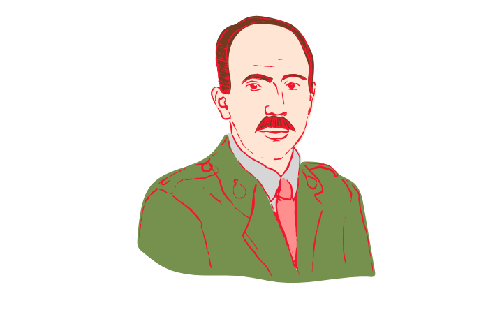
Born in New Zealand in 1882, Harold Gillies was a surgeon who supported on the Western Front during World War I. During his time on the Front, Gillies saw the devastating effects of the war, including the horrific facial injuries many soldiers suffered. He decided to use his skills as a surgeon to help do something about it, and became known as the ‘Father of Modern Plastic Surgery’.
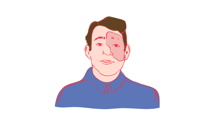
In 1917, Gillies convinced the army that they needed a hospital specifically for repairing facial injuries. So, he set up Queen’s Hospital in Sidcup, Kent. By sending postcards to base hospitals (see The Chain of Evacuation), Gillies invited soldiers who had suffered injuries to the face to come and be treated at his hospital. Him and his team carried out facial reconstruction surgery, often experimenting with new techniques like skin grafts.
It didn’t always go completely to plan, and could sometimes lead to the death of a patient. However, after trying different techniques, Gillies arrived at what became his signature method. He would take pieces of skin that were still attached to the body and graft them onto the face so there was still a blood supply. By the end of World War II, Gillies had treated 5,000 soldiers from across Britain, Australia, New Zealand and Canada. Afterwards, Gillies was made a knight for his contributions.
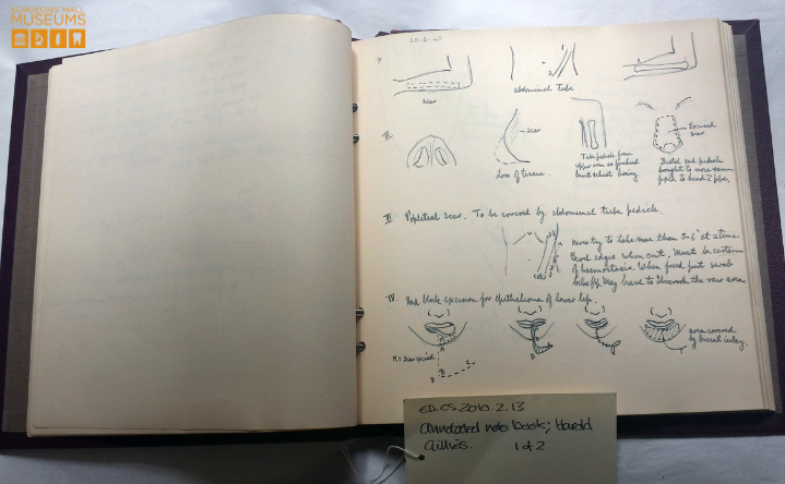
Take a look at these sketches done by Harold Gillies, which show us the complicated details of facial reconstruction. He wrote in this notebook during the Second World War at Rooksdown House in Basingtoke, which was a hospital wing that was converted into a plastic surgery unit to meet the needs of the war. You can view this notebook up close in our Source Gallery, where you can see some of Gillies’ ground-breaking techniques in detail.
Blood Transfusions
In 1900, a man named Karl Landsteiner made a discovery that blood belonged to four different types, and that only some of these types could be mixed. Until then, people had no idea that a patient losing blood needed to be supplied with blood from the same blood group, so carried out blood transfusions which ended up killing patients rather than saving them.
As you can imagine, blood was in huge demand during World War I. Knowing the different blood groups and how to mix them safely made blood transfusions possible, which were usually carried out at Casualty Clearing Stations. However, storing blood was tricky as it would clot quickly (which made it unusable). That was until Richard Lewisohn, an American scientist, found that blood could be mixed with sodium citrate to stop it clotting in 1914. An American army doctor named O. H. Robertson then tried this out during the war and found it kept blood fresh, which he used to save British soldiers. Later, it was discovered that refrigerating blood would also keep it fresher for longer and in 1917, a British surgeon named Geoffrey Keynes invented a machine that could transport blood safely.
Altogether, these developments made it much easier for transfusions to take place. The development of safe and effective blood transfusions saved thousands of soldiers on the Western Front, and meant countries were more prepared for blood transfusions when World War II broke out.
X-rays
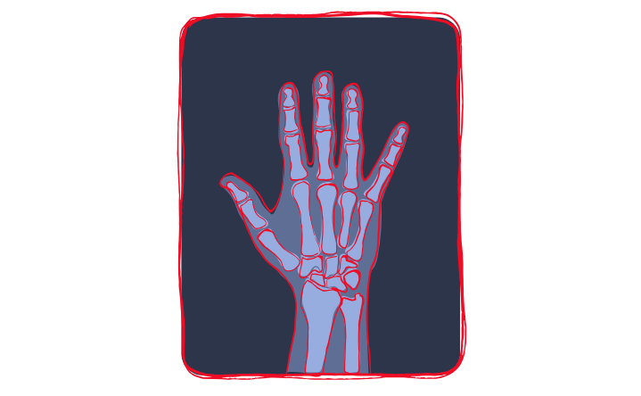
X-rays were first invented in 1895 by William Rontgen, and allowed doctors to see bones and diagnose injuries that couldn’t previously be seen, like breaks or fractures. However, they weren’t originally portable enough for use in World War I.
Marie Curie played a huge part in bringing X-rays to the battlefield. She left her work behind to create the first ever portable X-ray machine that could be used in the war. Her work meant that by 1916, X-ray machines were available at all of Britain’s Casualty Clearing Stations (CCSs). Though they weren’t perfect and didn’t work 100% of the time, they helped save lots of lives.
X-rays not only showed images of bones, but could also show the location of shell fragments and bullets in a soldier’s body. Often, soldiers died from these injuries because surgeons were unable to locate the bullet or shrapnel and so the wound became infected or bled out. X-rays meant that these foreign objects could be easily found and removed by surgeons more quickly, making it less likely for the soldier to become infected and lose blood.
Did You Know?
X-rays were first discovered by accident!
Amputation and Artificial Limbs
Because of the conditions at the Front, surgery wasn’t carried out in the clean and safe environments we see today. Though Joseph Lister’s ideas for hygienic best practice in surgery came before the war, there was no choice when it came to dealing with injuries in these circumstances. So, because of the dirty conditions, lots of soldiers had to have their limbs amputated to prevent infection and gangrene spreading. And because of the increasing number of amputations taking place, World War I led to improvements in artificial limbs, which are continuing to develop today.
Thomas Splint
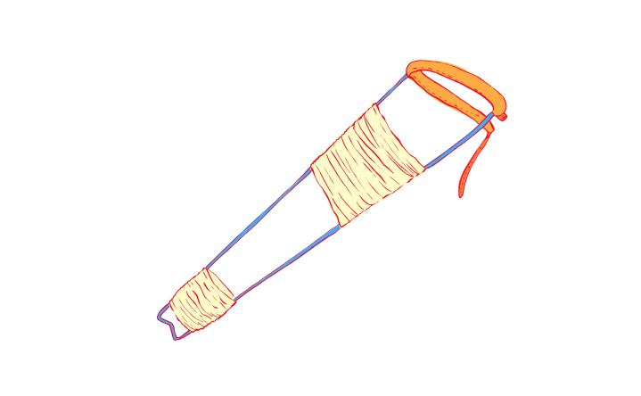
There were countless ways a soldier could be injured in World War Two, but a compound fracture was one of the most dreaded kinds of injury. It happens when a bone breaks and breaks through the skin, creating an open wound. Ouch! When compound fractures took place out on the battlefield, it wouldn’t be long until it became infected. In fact, it was the number of deaths from compound fractures of the femur (the thigh bone) that led to the Thomas splint being introduced.
It was originally designed by Hugh Thomas, a Welsh surgeon who gave the splint its name. The Thomas splint works by stabilising the fracture, which prevents both infection and blood loss. Thomas’s nephew, a surgeon named Robert Jones, saw his uncle’s design and brought it to the Western Front. This reduced the death rate of compound fractures from 80% to 20%. You can view a Thomas Splint that belongs to Surgeon’s Hall Museum in our Source Gallery.
Did You Know?
Variations of the Thomas Splint are still used today!
Quiz yourself!
Want to share feedback on this content? Get in touch or fill out the form.

